We have started our services for limited time. In case you are unable to come for follow up / check up
please contact us on +919822052063 / +919130025349/ +919130025350
Incase you have any difficulty for travel please let us know. We can send you a request letter.

We specialize in the diagnosis and management of complex eye diseases, offering state-of the-art infrastructure, surgical facilities, while at the same time providing routine eye care. Our team of specialists ensure that our patients get access to the most advanced treatment available in the world for all eye related problems. - Dr. Prabhudesai
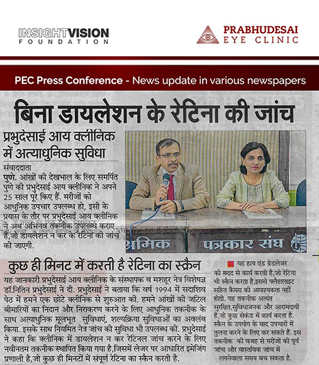
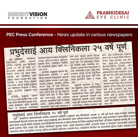
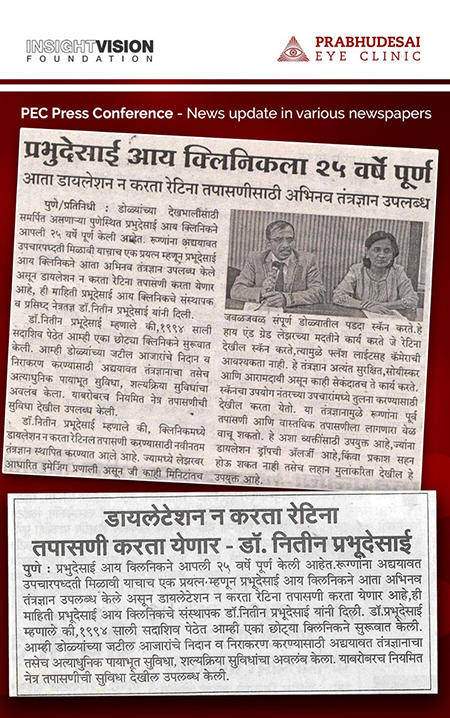
Prabhudesai Eye Clinic was started as a small eye clinic in the year 1994 in Sadashiv Peth Pune. We have successfully completed 25 years of dedicated eye service. In these years we have strived to offer latest, advanced and proven solutions to all our patients. We are committed to giving utmost care to the health and well-being of eyes of patients in this ongoing journey. New technologies have added for the treatment of eye diseases particularly for management of Retinal diseases and Glaucoma. Presently we offer top of the line instrumentation for the management of most eye diseases.
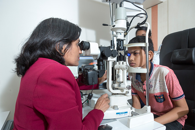
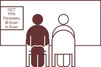
Retinal diseases are first evaluated clinically with Indirect Ophthalmoloscopy and Slit Lamp Biomicroscopy. Then, if needed, advanced tests are performed.
The advance tests include:
- Fundus Fluorescein Angiography: examination of the retinal blood vessels in detail.
- Optical Coherence Tomography: examination of the cross sectional structure of the retina.
Specialised treatment of retinal diseases include:
- Green Laser Photo Coagulation
- Red Laser Photo Coagulation
- Retinal Cryopexy
- Pneumoretinopexy - Gas injection
- Scleral Buckle Surgery for Retinal Detachment
- Vitrectomy with injection of Gas / Silicone oil for complex Retinal Detachment
- Indirect Laser Ophthalmoscope delivery system for treatment of premature babies affected by Retinopathy of Prematurity.
It is one of the commonest problems affecting vision in people having chronic diabetes where blood sugar is not well controlled.
Diabetic retinopathy leads to swelling of the central part of the retina, resulting in blurring of reading vision. If neglected it forms abnormal blood vessels on the surface of the retina which subsequently either bleed in front of the retina or cause pull on the retina. This ultimately results in profound loss of vision. Hence, every diabetic patient should have a periodic examination of his eyes and retina in particular to diagnose this problem at early stage to prevent major vision threatening problems later.
When a person undergoes thorough evaluation and is found to have Diabetic Retinopathy he usually undergoes tests like FFA (Fundus Fluorescein Angiography), OCT (Optical Coherence Tomography) and Perimetry. Based on these tests, the decision is taken on whether to go for Laser Photocoagulation or Intravitreal injection of Anti VEGF. In case of Laser treatment, it may be enough to go for a limited treatment of a single sitting. But if the problem is more serious, it may require extensive treatment with three to four sittings. In case one needs Anti VEGF injections, these injections are given on monthly basis till the problem is controlled. But even after the prescribed injections are over, periodic examination is needed keep a check on Retinopathy. At times, there may be a need for additional injections or Laser treatment. If it is a case of severe Retinopathy with bleeding in the eye or significant pull on the retina, then surgery is recommended in the form of Vitrectomy and intra operative Laser with injection of Gas or Silicone oil
![]() Watch Diabetic Retinopathy Video
Watch Diabetic Retinopathy Video
ARMD is a common cause of vision impairment in the elderly. It usually affects one after the age of 60.Commonly two types of problems are noted: Dry type and Wet type.
ARMD usually affects the central part of the retina, hence reading, writing, recognizing faces becomes difficult.
In the Dry type, progression of the problem is very gradual and presently no active treatment is available to halt the progression.
In the Wet type, the drop in vision is sudden due to swelling and bleeding at the centre of the retina. If left untreated, this problem can get serious. If ARMD is suspected after examining the retina, tests such as FFA (Fundus Fluorescein Angiography), OCT (Optical Coherence Tomography) and sometimes ICG (Indo Cyanine Green) angiography are performed to confirm the diagnosis and decide the treatment strategy. Currently for Dry type, no active treatment is available to improve the condition. Antioxidant tablets are prescribed to delay the progression of this condition. For the Wet type, as the condition worsens rapidly, treatment in the form of Injection of AntiVEGF drug into the eye is advised. Usually a loading dose of three injections on a monthly basis is given followed by injections as and when required. These injections are given with sterile precautions and are almost painless. In fact, one can be back to routine within an hour.
Retinopathy of Prematurity is a condition that affects premature infants which, if left untreated, can lead to profound loss of vision. Premature babies born before 32 weeks of pregnancy, with a birth weight of 1500 grams or less, are at risk of developing this problem. These babies need screening of the eye by 30 days post birth, to be continued up to 60-90 days on periodic basis.
If ROP is noted, then a close follow up is needed. If the disease progresses further, then to control it Laser Photocoagulation is applied. This can be performed under the effect of anaesthetic drops without causing much discomfort to the baby.
Post Laser Photocoagulation, the baby is examined periodically till the disease process comes under control. Even after control, these babies are kept under observation till their first birthday.
Retinal Detachment is an ocular emergency. Mainstay of treatment is surgery. Surgery involves supporting the Retina from outside with a Silicone band, applying Cryo to the Retinal defects and draining the sub retinal fluid. The complete procedure is done under local anaesthesia. Following surgery eye is patched and individual can go home after resting for a period of one hour.
![]() Watch Retinal Detachment Surgery Video
Watch Retinal Detachment Surgery Video
In conditions like Diabetic Retinopathy, hypertensive retinopathy our eye injury, bleeding can develop the eye. Rarely the bleeding can clear spontaneously but if it persists surgery in the form of Vitrectomy may be needed. During Vitrectomy Three 23/25 gauge instruments are introduced in the eye through the white part of the eye called sclera. The vitreous along with the admixtured blood is cut and sucked out of the eye. Once the bleeding is cleared the abnormal tissue is removed from the eye and the pull on the retina is relieved. Retina if detached is placed back to its normal position. SF6/C3F8 gas or Silicone oil is injected in the eye.
Post operatively the individual needs to position himself for a week to 10 days to get maximum benefit from the gas/oil . The gas gets absorbed on its own whereas the silicone oil needs to be removed surgically after three to six months.
![]() Watch Vitrectomy Surgery Video #1
Watch Vitrectomy Surgery Video #1 ![]() Watch Vitrectomy Surgery Video #2
Watch Vitrectomy Surgery Video #2 ![]() Watch Vitrectomy Surgery Video #3
Watch Vitrectomy Surgery Video #3
Older indivisuals at times develop a peculiar problem called Macular Hole where by a defect develops in the centre of the retina leading to loss of central vision. This problem does not have any medical treatment and surgery in the form of Vitrectomy with Gas injection is needed to treat this problem. Following surgery a face down positioning is required for a period of a week to 10 days.
![]() Watch Macular Hole Surgery Video
Watch Macular Hole Surgery Video
Glaucoma is a common vision threatening disorder of the eye that usually goes unnoticed in most cases till significant irreversible damage has already occurred. Awareness about this disease helps in early detection and prevention of major damage.
Though Glaucoma can rarely occur at birth, it is commonly seen after 40 years of age. Age, diabetes, high blood pressure, family history, smoking are the main risk factors for glaucoma after 40 years of age.
The primary problem in glaucoma is the damage of the main nerve of the eye- the Optic nerve. A common associated problem is increase in the eye pressure and decrease in the visual field.
Once the doctor suspects presence of glaucoma, he prescribes a group of tests called glaucoma work up to confirm the diagnosis. If the diagnosis is confirmed, the patient is prescribed anti glaucoma drops. Some may require up to three types of drops to control the progression of the disease process. Once started, drops are to be continued for life. If the drops fail to control the disease then the surgical option is considered.
A patient suspected to have Glaucoma or already diagnosed with the problem, needs to get a number of tests termed Glaucoma Workup. The tests include:
- Automated Perimetry
- Pachymetry
- Optic Disc photography
- Optical Coherence Tomography
- Oculyzer
- Specular Microscopy
Following diagnosis, there are several options available to treat Glaucoma:
- Medical management
- Laser Photocoagulation: - Laser Iridotomy with YAG Laser
- Surgical management: - Standard Drainage procedures or Advanced Glaucoma Drainage devices/implants
- Trabeculectomy: This is a standard Glaucoma surgery. Apiece of tissue in is removed, creating an opening. The opening is partially covered with a flap of tissue. This new opening allows fluid aqueous humor (aqueous humor) to drain out of the eye.
- Express Implant - Glaucoma Filtration Device: This device is an option to the routine Trabeculectomy surgery. This device increases the predictability and safety of glaucoma surgery.
- Ahmed Glaucoma Valve: This is yet another innovation in valve surgery used in complicated Glaucoma situations where there is a high chance of failure of routine Trabeculectomy surgery. Based on the valve principle, the implant drains excess fluid accumulating in the eye into the space surrounding the eyeball and stops draining once the pressure is normalised.
- Paediatric Glaucoma Surgeries: At times babies may be born with Glaucoma and may need immediate surgery to prevent permanent loss of vision. Using a special type of lens and high magnification of the operating microscope a procedure called Goniotomy is performed to clear the abnormal tissue and create a path for internal fluid flow. Other procedures like Trabeculectomy with Trabeculotomy are also performed to create a path for internal fluid flow. Even after surgery, these infants need regular check-ups as a lifelong safeguard against Glaucoma.
Cataract is a common problem of old age. In this condition the normal crystalline lens of the eye starts getting cloudy, resulting in blurred vision. This leads to difficulty in day to day activities as reading, writing, watching TV, recognising faces at a distance, reading signboards from a distance etc.
Though age is the main cause of cataract, diseases like Diabetes, High Blood Pressure, High Cholesterol and habits like smoking, chewing tobacco, drinking can hasten the onset of cataract.
Certain eye conditions like chronic inflammation of the eyes, retinal detachment, glaucoma, long term use of steroid drops, injury to the eye, increases the chance of cataract development at an early age.
Once the cataract sets in there is no medical treatment for it. Treatment is usually in the form of surgery which involves removal of the cataractous lens and implantation of an artificial lens.
Cataract surgery involves removal of the natural lens of the eyes which becomes opaque and hard, blurring vision. We perform advanced cataract surgeries including Paediatric cataract surgery. A choice of premium intraocular lenses is available for patients. If both eyes need surgery, it can be planned within a span of few days.
The standard procedure is Phacoemulsification. A small pen like probe is introduced in the eye and using ultrasonic energy, the hard part of the cataract called Nucleus is emulsified or broken down into very fine pieces and sucked out of the eye. Then the remaining soft part is just sucked out of the eye. Once the cataract is cleared then an Intraocular lens is introduced in the eye in a folded manner through the original opening. This lens then unfolds in the eye.
As the wound is small, it heals fast. The patient can get back to his routine the next day itself. However, as an added precaution, it is advisable to avoid hectic activities and outdoor jobs for the first week.
Use of Intra ocular lens is now standard of care for Cataract surgery. The commonest type of Intra ocular lens is called as unifocal lens which focuses images of distant objects on to the back of the eye and hence glasses are needed only for reading. The other type of lens is called multifocal lens which focuses the distant, intermediate and near objects on the back part of the eye. Hence you do not need glasses for most common activities.
Premium Intraocular Lens
- Toric Intraocular Lens for correction of astigmatism.
- Multifocal Intraocular Lens for correction of near, intermediate and distant vision.
- Multifocal Toric Intraocular Lens for correction of astigmatism and near, intermediate and distant vision.
LASIK is a type of refractive surgery done with the help of Laser which helps to get rid of Refractive errors which in simple terms means to get rid of glasses and contact lenses.
We usually have three types of refractive errors of the eyes:
- Myopia or Short-sightedness
- Hypermetropia or Long-sightedness
- Presbyopia or difficulty in near vision after age 40
LASIK is usually advised when the glass power is stable for about a year. This is usually seen after completion of 18 years of age.
Pre-Treatment Tests for LASIK SurgeryIf an individual is interested in having Excimer Laser treatment he needs to undergo certain tests like
- Advanced corneal topography with Oculyzer machine
- Corneal endothelial cell count with Specular microscope
- Corneal measurements with Optical Biometer
Based on these reports it is decided whether the individual is fit for Excimer Laser. If fit the procedure is performed on day care basis, with the help of EX 500 Excimer one of the fastest laser available for this purpose. This makes the treatment short, precise and comfortable.
The main procedure requires about 5-10 minutes. No eye patch is required hence the person can leave the clinic in a few minutes after the procedure is done. Usually the individual can resume his duties after a short rest of 2-3 days.
Cornea is the front most transparent part of the eye. It is often referred to as ‘black of the eye’.
Corneal Diagnostics: - for precise diagnosis of corneal conditions we have various tests like a. Oculyzer , b. Aladdin topographer, c. Topcon Specular Microscope, d. Pachymeter, e. Photo slit lamp, etc.
Various disorders affecting the cornea can lead to pain, irritation, reduced vision, and sometimes blindness.
The most common of these disorders are:- corneal infections, corneal scars – post trauma or infection, immune mediated disease, keratoconus, chemical injuries etc.
Treatment options for all of them are now available. They include corneal transplant, lamellar corneal surgery and stem cell transplant and therapy, where the diseased cornea is removed and replaced with a natural cornea procured from a donor eye (eye donated after death). Artificial cornea is also available now, for cases where multiple corneal transplants have failed.
Other than corneal surgery for corneal diseases, we conduct refractive surgery for reduction of refractive error (glass number) and phakic intraocular lens implant surgery to correct myopia.
Keratoconus treatment we offer C3R therapy. For Dry eye problem we offer complete work up and treatment including punctual plugs.
Misalignment of both eyes is called strabismus or squint. There is loss of coordinated movements of both eyes either due to ocular muscle imbalance or their nervous control.
It is usually present in childhood but may develop in adults as well. The squint may be constant or intermittent. Some children may close one eye in bright sun light or acquire abnormal head posture.
Vision in both eyes may be normal but may be less in squinting eye due to amblyopia (lazy eye).There is loss of binocular vision and stereopsis (depth perception).
- Assessment of strabismus
- Refraction (spectacle correction)
- Ocular movement testing
- Binocular vision testing
Treatment: aims at restoring good vision in each eye, alignment of both eyes and binocular single vision.
It may be in the form of
- Spectacle correction
- Occlusion therapy for amblyopia (lazy eye)
- Muscle exercises
- Surgical correction
Ophthalmic plastic surgery is a specialized area of ophthalmology dealing with cosmetic and functional deformities and abnormalities of the eye. It includes drooping of lids, defectsin the lacrymal system, the orbit and the periocular region.
Oculoplastic Surgery is plastic surgery as related to the area around the eye. The eyelids and surrounding areas are extremely complex and delicate structures that are vital to the preservation of sight and are important features of a normal facial appearance. Since surgery in this area can affect one’s ability to see, Oculoplastic Surgeons are uniquely qualified to perform this delicate surgery and to provide care to the eye if needed. This service takes care of drooping of lids, lid scars, watering of the eyes, thyroid related eye problems etc. Additionally we also offer Botulinum injection for facial spasms.
Various types of lasers are used in eye care. At our clinic we offer A. Green laser for Retinal disorders, B. YAG Laser for post cataract and glaucoma problems, C. Excimer laser for removal of glass power for retinal problems. These treatment can be performed in the out-patient department or in the OT depending on the problem. Laser treat can be performed without giving injection and one can get back to work the next day itself.
Uveitis is a chronic disorder involving swelling and irritation of the Uvea uvea, the middle layer of the eye that provides most of the blood supply to the Retina retina, if not treated properly, it can lead to significant loss of vision. However, timely evaluation, investigations and management with specific drugs can control it. Also surgical procedures like intravitreal injections of steroids / Anti VEGF agents & Vitrectomy surgery are performed in case of complications.
“Low” means not normal and “Vision” means person is not blind.
Most individuals today classified as "blind" actually have remaining sight and, thanks to developments in the field of low vision rehabilitation, can be helped to make good use of it, improving their quality of life. It affects rich and poor, old and young, all races and ethnic backgrounds. It has been estimated that by 2020, 15.5% of the population will be older than 65 years and will include an increasing number of potential low vision patients.
Many visually impaired people experience difficulty with regular activities such as cooking, shopping, managing finances, watching television, reading, and taking care of their personal needs. Many people with low vision have concerns about finding and keeping a job, dealing with friends and family members, getting around and participating in recreational and social activities.
Knowledge is the key to living with low vision. People with low vision can enhance their quality of life by learning which optical and non-optical devices can help them. Low vision specialists recommend appropriate low vision devices and counsel patients on how better to deal with their reduced vision in general. But knowledge is just the first part. Practice is the important next step. Just as an amputee must learn to walk with his new prosthesis, a person with low vision must practice using adaptive aids until they become second nature. Motivation and attitude are perhaps the most important factors in determining one's success in using low vision devices. If people are willing to give some energy and effort to learning how to make the best use of their remaining sight, they can do nearly everything they did before, perhaps just in a different way.
We have a well equipped clinic and trained specialists to render the services to patients with low vision. Patients with Macular Degeneration due to ageing or other factors need vision enhancement of with the help of special visual aids. This service offers such aids after proper evaluation of the patients’ needs and lifestyle.
A contact lens is a synthetic lens that sits on the surface of the eye and helps correct vision problems like long-sightedness, short-sightedness, presbyopia and astigmatism
Most contact lenses fall into one of two groups:
- Soft lenses made of a very soft and flexible, water-absorbing material that conforms to the eye shape
- Hard lenses made of rigid gas-permeable plastic that cover only part of the cornea.
Contact lenses can fill almost any vision correction need including myopia (main use), hyperopia, astigmatism and presbyopia. Contact lenses also have certain therapeutic uses and are needed for bandage lenses, or even in some cases for drug delivery. Opaque contact lenses may be used to change eye colour or to hide disfigurement of the cornea such as scars.
Contact lenses can be classified according to:
- Function:
- Material:
- Replacement Schedule:
- Spherical contact lenses come in the typical, rounded design, which can correct myopia (nearsightedness) or hyperopia (farsightedness).
- Toric contact lenses are the correct choice for astigmatism, as well as for myopia and hyperopia
- Bifocal contact lenses contain different zones for near and far vision, to correct presbyopia
Corrective: Lenses to correct refractive disorders of the eye.
Therapeutic: Lenses for the treatment of non-refractive disorders of the eye.
Cosmetic: To change the appearance of the eye.
Soft lenses: Contact lenses that are manufactured from plastic materials that are able to absorb water, also called "hydrogels." They range from low oxygen permeability to high oxygen permeability (silicone hydrogels).
Rigid Gas Permeable Lenses (GP Lenses): Contact lenses that are manufactured from plastic materials that have high oxygen permeability and do not contain water.
Conventional: No prescribed replacement schedule. The contact lens is replaced every six months or more or when it wears out, becomes damaged, or the patient's power requirements change.
Planned: There is a specific time period after which the contact lens will be replaced.
Disposable: A contact lens that is disposed of upon removal from the eye and replaced.
Gas permeable contact lenses are more resistant to lens deposits and don't need to be discarded as frequently as soft lenses. Often, GP lenses can last a year or longer before they need to be replaced.
Many lens designs are available to correct different types of vision problems:
Premature infants, especially those with birth weight less than 1500 grams are at risk of developing a retinal problem called Retinopathy of Prematurity. This problem can be detected and controlled if the baby is periodically examined as advised by the Paediatrician in consultation with an Eye specialist. Also babies can have certain birth defects or diseases like glaucoma at birth which may need immediate treatment to prevent a major visual problem.
Any white reflex in the eyes of children below 5 years needs a thorough medical check-up as it could be a sign of a major eye problem. Similarly, any eye with constant pain, redness, watering needs proper check-up and appropriate treatment. Children with squint also need thorough check as in many cases it is possible to correct the squint surgically.
In this age group, the correction of lens power is of prime concern. Use of glasses, contact lenses and correction of the lens number with the help of Laser are the available options.
For those over 40 years of age, correction of near vision is required. At this age a comprehensive eye check is needed to pick up early vision affecting diseases.
Individuals with high minus number (myopia) need to have their retina checked on an yearly basis. Individuals with Diabetes, Blood pressure, High Cholesterol or any other systemic illness like Tuberculosis and HIV need to have their eyes checked up regularly as in these conditions there is high chance of vision getting affected.


- Topcon Swept Source 'Atlantis' OCT DRI OCT System (Non-Invasive Angiography)
- Topcon Digital Angiography System
- Topcon Aladdin Optical Bio-meter with Topographer
- Topcon Specular Microscope
- Alcon Infinity Phacoemulsification System
- Alcon Wavelight Oculyser Advanced Topographer
- Alcon Wavelight EX 500 Excimer Laser
- Constellation Vision System
- Leica M 822 Ophthalmic Microscope with HD Camera
- Cirrus OCT from Zeiss
- Lumera I Surgical Microscope from Zeiss
- Humphrey Visual Feild Analyser
- HD Camera for Watching and Recording Live Surgery
- YAG Laser from Lightmed
- Alcon Pure Point Green Laser
- Nidek Green Laser
- Medivision Slit Lamp Camera
- Nidek Ultrasonography Machine
- Flash Autoclave
A pioneer in introducing technological innovations in the city, Prabhudesai Eye Clinic has certain firsts in Pune:
Digital Angiography system allows high quality Fundus Fluorescein Angiography, a test performed for examination of the retinal blood vessels in diseases like Diabetic Retinopathy. With the hi- tech system, the test is performed rapidly and precisely helping the doctor with correct diagnosis and treatment.
Optical Coherence Tomography allows us to scan the central retina which helps in diagnosis of diseases like Age Related Macular Degeneration, Diabetic Retinopathy, Venous Occlusions & CSR to name a few.
Constellation Vision System is a combined surgical system which is used to perform complex Vitreoretinal surgeries and Hi tech Cataract surgery. This is one of the most advanced surgical systems currently available internationally, with potential for future upgrades when needed.
Leica Microscope, an advanced microscope with dual illumination for Cataract as well as complex Vitreoretinal surgeries. This recently launched microscope allows precise and perfect surgeries allowing rapid and easy recovery for the patient.
Prabhudesai Eye Clinic was started as a small eye clinic in the year 1994 in Sadashiv Peth Pune. Step by step new technologies were added for the treatment of eye diseases particularly for management of Retinal diseases and Glaucoma. Presently we offer top of the line instrumentation for the management of most eye diseases.
Prabhudesai Eye Clinic is committed to shaping the vision of providing eye care of international standards at affordable costs. We seek to achieve this by continuous upgradation of knowledge and technology. At the same time, we focus on the training of eye care providers who share our mission to reach out to the broadest cross section of society with our service offerings.
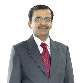
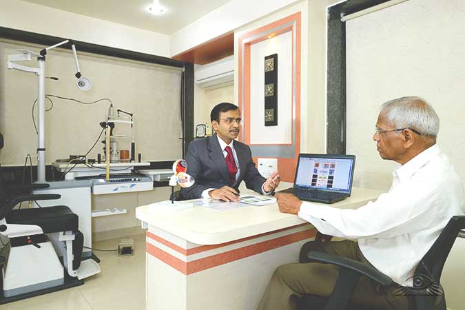
M. S. (BOM.) D. O. M. S.,
Vitreo Retinal Surgeon
Dr Prabhudesai , an alumni of TN Medical College-Nair Hospital Mumbai, had his super speciality training in medical and surgical management of Vitreoretinal disease at the prestigious Shankara Nethralaya Chennai. Subsequently he worked as a Vitreoretinal consultant at the same institute, followed by a stint at Shri Ganapati Netralaya Jalna.
Since 1994 he has set his own practice in the city of Pune. In 1998, he founded Prabhudesai Eye Clinic, along with his wife, Dr Medha Prabhudesai. The clinic has been steadily growing over the years and presently offers service for Vitreoretinal disorders and Glaucoma on par with international standards. Inspite of his busy private practice Dr Prabhudesai is actively involved in academic activities. He is an active member of Poona Ophthalmological Society, Maharashtra Ophthalmological Society, All India Ophthalmological Society & is founder member of Retina Interest Group Pune. He is also an International member of the prestigious American Academy of Ophthalmology.
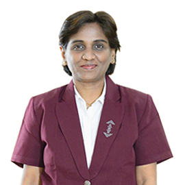
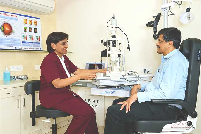
M. B. B. S., D. O. M. S,
Cataract & Glaucoma Surgeon
Dr Medha ,an alumni of B.J Medical College, Sassoon Hospital Pune, completed her super speciality training in medical and surgical management of Glaucoma & Anterior segment disorders of the eye at the prestigious Sankara Nethralaya, Chennai. She followed her training with a stint as a Glaucoma and Cataract consultant at Shri Ganapati Netralaya, Jalna.
Currently she is practising as a Glaucoma consultant at Prabhudesai Eye Clinic a post she held since 1994. Dr Medha has quite a few significant milestones in her career. For instance, she initiated the process that established Glaucoma as a sub speciality in the city of Pune. She is,in fact, a founder member of Glaucoma Interest Group, Pune.
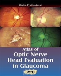
Her passion for her subject led her to complete five studies on various anti- glaucoma molecules as well as publish a book titled: Atlas of Optic Nervehead Analysis in Glaucoma (available on the international medical circuit). Also, her video titled Bleb Repair using Scleral Autograft was presented at the annual conference of the prestigious American Academy of Ophthalmology at Orlando in Oct 2011.
While continuing her private practice, Dr Medha is also an active member of Poona Ophthalmological Society, Maharashtra Ophthalmological Society & All India Ophthalmological Society.
Through the Trust we try to help the economically weaker sections of society to undergo specialized treatments. We try our best to accommodate all patients referred to us. Any donations made towards the Trust are welcome and are utilised only for patient care. The Trust also undertakes activities like training programmes for doctors and paramedical personnel.
We have introduced a division ‘Spectra Vision Care’ that will cater to refractive surgery, popularly known as LASIK, which will help individuals in getting rid of their glasses or contact lenses. This procedure is best suited for anyone over 18 years of age with a stable glass power for a period of (approximately) one year.
As part of the screening process these individuals would first undergo a diagnostic test to ensure that they are suitable for the treatment. After verification, the patient can undergo the surgery with the most advanced technology, namely EX 500 Eximer Laser, Wavelight Germany, by Alcon, the world leaders in ophthalmic instruments. We assure that with this technology we will provide fast, safe and predictable results.
Typically the main laser treatment takes a few seconds, and the overall time in the Laser suite would be just a few minutes, so the in and out time in the clinic would be about an hour. The overall recovery time from this procedure is short, so if one gets the surgery done by Friday evening, then he/she can get back at work by Monday morning, with only post-surgery eye drops required over the next few weeks.
We are happy to hear from you, please contact us for further information.
We have introduced a new division ‘Spectra Vision Academy’ that will cater to educating interested 10th/12th pass students in the field of ophthalmology.
The course offerings are:
- Diploma in Operation Theatre Technology - 2 years
- Diploma in Optometry - 2 years
- Eye Technician - 1 year
- Certified Ophthalmic Assistant - 1 year
- Diploma in Ophthalmic Assistance - 1 year
Dr. Nitin Prabhudesai and Dr. Medha Prabhudesai where invited to MOS Annual Conference.
Dr. Nitin Prabhudesai gave talks on three topics i.e. “Role of Bio-Similars in Retinal Disorders”, “Paediatric Retinal Disorders” and “Post-Operative Endophthalmitis Management”
Dr. Medha Prabhudesai gave talks on three topics i.e. “Role of Optic Disc Evaluation in Glaucoma Management”, “Scleral fixation IOL” and “Pre Perimetric Glaucoma”. Aurangabad - 26th to 28th October 2018.
Dr. Medha Prabhudesai was invited to give a talk on "Medical Management of Glaucoma" at the Annual Conference of GSI. Thiruvananthpuram - 14th to 16th September 2018.
Dr. Nitin Prabhudesai was invited as a panellist for ‘NVision Education Retina’, a discussion forum arranged by Novartis India. He shared the platform with international speaker Dr. Patricio Schlottmann from Argentina, where various Retinal topics of interest were discussed. JW Marriott Hotel, Pune - 4th September 2018.
Dr. Nitin Prabhudesai was invited as a faculty for the Annual Conference of the Bombay Ophthalmologist’s Association and gave talks on ‘Management of Recurrent Vitreous Haemorrhage’ and ‘Will Biosimilars reduce the use of Off Label drugs in Ophthalmology?’
The talks were well received by delegates and panellists which lead to healthy discussions and awareness of the topics. Mumbai
- 24th to 26th August 2018.
Prabhudesai Eye Clinic conducted a well-attended & appreciated CME (Continuing Medical Education) program in “Refractive Surgery for All” at Siddharth Palace Hall, in front of Prabhudesai Eye Clinic, Pune, Kothrud - 12th August 2018.
Dr. Medha Prabhudesai was invited as a faculty for Glaucoma Webinar, an online program for ophthalmologists pan-India. She gave a talk on ‘Optic Nerve Head Evaluation’and had follow-up Q&A session with online participants - 8th August 2018.
Dr. Nitin Prabhudesai was invited as a faculty for the ‘Refractive Surgery Conclave’ by Narayan Nethralaya at Bangaluru - 4th to 5th August 2018.
Dr. Medha Prabhudesai participated in the Glaucoma Experts Meet, a gathering of national and international Faculty from India, Singapore and Australia. Mumbai - 3rd to 5th August 2018.
Dr. Nitin Prabhudesai gave a talk on ‘Paradigm shift with Biosimilar introduction in Ophthalmology’ at the Retina CME organised by Bombay Ophthalmic Association, Mumbai - 15 July 2018.
Dr. Nitin Prabhudesai was ceremoniously elected as the Honorary President of Poona Ophthalmological Society on 1st April 2018 event at Hotel President, Pune. He envisions his tenure from 1st April 2018 to 31st March 2019 as a humble opportunity to plan and execute educational activities for the members of this prestigious society. He also plans to extend the training programs to the postgraduate students of Ophthalmology, thus creating a new generation of well-trained Ophthalmologists.
Along with him, Dr. Aparna Vaidya took over as Honorary Secretary and Dr. Monika Naik Nimbalkar took over as the Honorary Treasurer.
Poona Ophthalmological Society has a 68-year legacy and is actively engaged in bringing to its members the recent advancements in ophthalmology. The society is also actively involved in conducting public awareness programs and encouraging its members to devote time to social service and public welfare - 1st April 2018.
Dr. Nitin Prabhudesai and Dr. Medha Prabhudesai participated in the annual conference of All India Ophthalmological Society (AIOS) held at Coimbatore between 22nd and 25th February 2018.
Dr. Medha Prabhudesai was instructor for instruction course on clinical examination in Glaucoma Surgeries, judge for Anterior Segment Video Session and moderator for free paper in Glaucoma.
Dr. Nitin Prabhudesai was chairman for free paper session in Vitreoretinal Disease and judge for various session on Posterior Segment Surgeries - 22nd to 25th February 2018.
We now offer a Non-Invasive OCT Angiography that uses the Swept Source OCT (Optical Coherence Tomography) technology instrument ‘Atlantis’ DRI OCT from Topcon, Japan, which allows faster and detailed evaluation of the back side of the Eye – Retina and Optic Nerve and evaluates the Retinal Vasculature without injecting the dye. In our endeavour to regularly upgrade to latest technology available in eye care, we are now amongst the few in Maharashtra to offer these services. - 9th December 2017.
Dr Nitin Prabhudesai and Dr Medha Prabhudesai participated in Annual Conference of Maharashtra Ophthalmological Society, which was attended by about 1500 delegates from all over Maharashtra. Dr Nitin Prabhudesai gave talks on 'Complications and Management of Macular Hole surgery', 'Cystoid Macular Oedema', 'Case report on Juvenile Retinoschisis' and 'Scleral Buckle - still the Gold standard in management of Simple Retinal Detachments'. He was the Chairman for two sessions on Vitreoretinal Surgery. Dr Medha Prabhudesai gave talks on 'Management of Allergies of Glaucoma edications' and 'Detecting Glaucoma progression on OCT'. She was a panellist for Glaucoma Grand rounds and Co-Chairwoman for the Instruction Course on Over Diagnosis of Glaucoma. The Conference was held at J. W. Marriot Hotel, Pune from - 13th to 15th October 2017 .
Dr Medha Prabhudesai gave a talk on 'Optic Nerve Head evaluation in Glaucoma', and participated in the debate on 'Medical v/s Surgical management of Glaucoma' at Annual conference of Glaucoma Society of India at Jaipur - 15th to 17th September 2017.
Dr Nitin Prabhudesai and Dr Medha Prabhudesai presented a poster titled 'Low incidence and less severe Retinopathy of Prematurity (ROP) at a tertiary care urban centre in Western India', at the Euretina 2017 conference Barcelona Spain - 7th to 10th September 2017.
Dr Nitin Prabhudesai attended the 'Aflibercept Adivisory Board Meeting', and moderated one session at Hotel Sahara Star, Mumbai - 3rd September 2107.
Prabhudesai Eye Clinic commemorated its 23nd Foundation Day by conducting a Continuing Education Program (CME) on 'Recent advances in Cataract Surgery', a well received and appreciated program, which was attended by prominent local Ophthalmologists at Siddharth Palace Hall, Kothrud, Pune - 20th August 2017.
Dr Nitin Prabhudesai gave a talk on 'Laser Treatment of Diabetic Maculopathy' at the Annual Conference of Bombay Ophthalmic Association in Mumbai - 11th - 13th August 2017.
Dr Medha Prabhudesai gave talks on 'Management of Angle Closure Glaucoma' and 'Management of Uveitic Glaucoma' at the conference on 'Decision Making in Glaucoma' under the aegis of Poona Ophthalmological Society and Maharashtra Ophthalmological Society, Pune - 30th July 2017.
Dr Nitin Prabhudesai attended the 'Global Retina Network Program' and had the opportunity to interact with world authorities in retinal diseases like Dr Peter Kaiser from USA and Dr Gemmy Cheung from Singapore at Seoul, South Korea - 8th July 2017.
Dr Medha Prabhudesai gave a talk on 'Imagining in Glaucoma' at the Glaucoma CME program held at H V Desai Hospital in Pune - 22nd Jan 2017.
Dr Nitin Prabhudesai and Dr Medha Prabhudesai were invited as faculty members for the Annual Conference of the Maharashtra Ophthalmological Society, an event attended by about 1500 delegates. Both of them gave three talks on Glaucoma and Retinal Diseases, they also judged the Free Papers Session. Dr Medha's talk on Neuroprotection, which was a part of special session by AIOS (All India Ophthalmological Society), was highly appreciated by the gathering at Vashi, Navi Mumbai - 14th to 16th October 2016.
Dr Medha Prabhudesai was a panellist in the Live Webinar Event on Glaucoma, where more than 1000 ophthalmologist from all over India logged in to participate in this event held by the prestigious All India Ophthalmological Society - 13th October 2016.
Dr Medha Prabhudesai conducted four interactive sessions on various aspects of Medical and Surgical Management of Glaucoma, as a guest faculty at CME on Glaucoma in Solapur - 15th August 2016.
Dr Nitin Prabhudesai and Dr Medha Prabhudesai attended the EURETINA 2016 conference at Copenhagen, Denmark - 8th to 11th September 2016.
Dr Nitin Prabhudesai gave a talk on 'Razumab - Real life experience' at CME by Intas in Pune - 31st August 2016.
Prabhudesai Eye Clinic commemorated its 22nd Foundation Day by conducting a Continuing Education Program (CME) on 'Retina for General Ophthalmologist' in association with Poona Ophthalmological Society at Siddharth Palace Hall, Kothrud, Pune - 21st August 2016.
Check the events post on Prabhudesai Eye Clinic's Faceook Page
Dr Nitin Prabhudesai gave a talk on 'Current concepts in management of Retinal Detachment' at the annual conference of Bombay Ophthalmological Society's annual conference in Mumbai - 20th August 2016.
Dr Medha Prabhudesai conducted a workshop on 'Multifocal IOL in day to day practice' in Pune - 15th August 2016.
Dr Nitin Prabhudesai gave a talk on Protocol-T in management of DME, at CME by Novartis in Pune - 7st August 2016.
Dr Medha Prabhudesai gave a talk on 'Congenital Glaucoma' at Maharashtra Ophthalmological Society's CME in Pune - 31st July 2016.
Dr Nitin Prabhudesai gave a talk on 'Combined Cataract and Vitrectomy Surgery' at Maharashtra Ophthalmological Society's CME in Pune - 31st July 2016.
Dr Medha Prabhudesai presented a talk on 'Glaucoma and Ocular surface disorders' on the invitation of Ahmednagar Ophthalmic Society and conducted an interactive session on Glaucoma at Ahmednagar - 27th March 2016.
Dr Nitin Prabhudesai conducted a course on 'Refractive Surgery' with Dr Nitin Balkrishnan as the guest speaker at Spectra Vision Care, Pune - 20th March 2016.
Dr Nitin Prabhudesai presented a talk on 'Evolving Ranibizumab Biosimilar, A Developmental and Clinical Review' at MAPROCON 2016, Hotel Ambassador Ajanta, Aurangabad - 7th February 2016.
Dr Nitin Prabhudesai presented a talk on 'Cytoid Macular Edema, Management of Diabetic Retinopathy with Cataract' at Netrakumbh 2016 - 1st Annual Conference of Nashik Ophthalmological Association, Jupiter Hotel, Nashik - 24th January 2016.
Dr Nitin Prabhudesai presented a talk on 'Cataract Surgery in AMD', also Dr Medha Prabhudesai presented 'Glaucoma Surgery in presence of Cataract' at POS Annual conference, Marriot Pune - 4th to 6th December 2015.
Dr Nitin Prabhudesai and Dr Medha Prabhudesai attended the AAO Annual conference at Sands Expo and Venetion at Las Vegas - 14th to 17th November 2015.
Dr Nitin Prabhudesai presented a talk on Anterior Vitrectomy, also Dr Medha Prabhudesai presented on 'OCT in Glaucoma' and 'Trabeculectomy Surgery - scientific basis of surgical steps' at the Annual Conference of Maharashtra Ophthalmological Society in Goa 23rd to 25th October 2015 .
Dr Medha Prabhudesai presented a talk on Imaging in Glaucoma at GSI Annual conference at Grand Hyatt Mumbai - October 2015.
Introducing the most advanced technology EX 500 Eximer Laser, Wavelight Germany, by Alcon, the world leaders in ophthalmic instruments, at 'Spectra Vision Care' for corrective vision - 25th September 2015.
Dr Nitin Prabhudesai attended the 8th AMD Congress at New Delhi - 8th and 9th August 2015.
Dr Nitin Prabhudesai and Dr Medha Prabhudesai attended the Sankara Nethralaya Alumni meet - 19th July 2015.
Dr Nitin Prabhudesai attended the Retina Vision day at Zurich – 26th to 27th June 2015.
Dr Medha Prabhudesai presented a talk on Imaging in Glaucoma, Glaucoma Update at Doctor Eye Institute at Mumbai - 14th June 2015.
Dr Nitin Prabhudesai and Dr Medha Prabhudesai presented a talk on Eye Care and Eye Diseases the Vasant Vyakhanmala, Tilak Smarak, Sadashiv Peth at Pune - 25th April 2015.
Dr Medha Prabhudesai presented a talk on Imaging in Glaucoma, at the DOS Annual Conference Hotel Ashoka in New Delhi - 10th to 12th April 2015.
Dr Medha Prabhudesai gave a talk on Ex-pres Glaucoma Device during the Glaucoma CME (Continuing Medical Education) held by Poona Ophthalmological Society at Pune - 2nd March 2014.
Dr Nitin Prabhudesai and Dr Medha Prabhudesai attended the 7th International Congress on Glaucoma Surgery at Singapore and presented their work on OCT in Glaucoma and Neovascular Glaucoma in poster presentation - 20th to 22nd February 2014.
Dr Nitin Prabhudesai participated in a round table discussion on management of Diabetic Retinopathy with world renowned retinal specialist Dr Neil Bressler & Dr Sussan Bressler from John Hopkins University USA at Mumbai - 28th January 2014.
We are happy to announce our new venture a “Premium Eye Care” service facility at the Ophthalmology Department of Ruby Hall Clinic, Wanowarie. - 9th January 2014.
Dr Nitin Prabhudesai and Dr Medha Prabhudesai were invited as faculty in Annual conference of Poona Ophthalmological Society held in Pune - December 2013.
Dr Nitin Prabhudesai and Dr Medha Prabhudesai were invited as faculty in the Annual conference of Maharashtra Ophthalmological Society held at Nagpur - October 2013.
Dr Nitin Prabhudesai attended the prestigious EURETINA Congress at Hamburg and interacted with world renowned retinal surgeons. - September 2013.
Dr Medha Prabhudesai gave a talk on 'Glaucoma in Uveitis' and Dr Nitin Prabhudesai gave a talk on 'White Dot Syndromes' during the Uveitis and Retina CME organised by Poona Ophthalmological Society at Poona Hospital - 1st September 2013.
Dr Nitin Prabhudesai presented an interesting case of Age Related Macular Degeneration in the 7th Indian AMD and RETINA Congress at Banglore - 17th August 2013.
Dr Nitin Prabhudesai delivered two talks in FOCUS-2013, The annual conference of Bombay Ophthalmic Associations
- Newer applications of SD-OCT in day to day practice
- Indications of Vitrectomy for Primary Retinal Detachment
at Mumbai - 3rd August 2013.
Dr Medha Prabhudesai gave a presentation on "End Stage Glaucoma" at Hotel Le Meridian - 23rd June 2013.
Dr Medha Prabhudesai gave a presentation - "Role of RNFL and RGC analysis on OCT for evaluation of Glaucoma" at Hotel Courtyard Marriot - 16th June 2013.
Dr Nitin Prabhudesai presented “Recent Advances in OCT” at Kashibai Navale Hospital, Pune, under P.O.S., CME 2013 - 10th March 2013.
Dr Nitin Prabhudesai presented “Retina Update” AFMC, under P.O.S. - 24th February 2013.
Dr Nitin Prabhudesai presented “Management of Retinal Venous Occlusions”, Hotel Pride. - 23rd February 2013.
Dr Medha Prabhudesai gave a presentation on “Medical Management of Glaucoma”, at the Annual Conference of Poona Ophthalmological Society, YASHDA, Pune - December 2012.
Dr Nitin Prabhudesai attended the World Retina and AMD Congress at Prague. - November 2012.
Dr Medha Prabhudesai gave a presentation on “Medical Management of Glaucoma”, at the Annual Conference of Maharashtra Ophthalmological Society, Mahabaleshwar - October 2012.
Dr Nitin Prabhudesai presented paper on “Management of Wet AMD” at 6th AMD Cogress at Dubai - July 2012.
Dr Medha Prabhudesai started Glaucoma Club for individuals having Glaucoma to create awareness about the disease - 16th March 2013.
Dr Medha Prabhudesai attended the “Frankfurt Retina Meet”, and subsequently attended Dr Ekhardt's Clinic, Frankfurt - March 2012.
Dr Medha Prabhudesai presented Instruction Course on OCT in Glaucoma, at the Annual Conference of Glaucoma Society of India, Coimbtore - 2012.
For first timers, comprehensive eye check is done to look for possible problems. The complete procedure takes about 1.5 to 2 hours. Hence it advisable that one takes a proper appointment and comes prepared to avoid unnecessary waiting.
In case of conditions like chemical injury to eye, acute loss of vision, severe pain in the eye appointment may not be needed between 8 am-8 pm.
A comprehensive eye check which includes history taking, glass power check, Slit lamp examination followed by dilated retinal evaluation and discussion with the patient requires approximately two hours.
Note: Diabetic patients should consider their food timings before fixing appointment.
It is preferable not to drive for about an hour after dilated eye exam.
A yearly checkup after the age of 40 years is enough. However if the patient has Diabetes, Blood pressure or any other illness then the frequency of visits will be decided after discussions with the Doctor.
Only one adult needs to accompany the patient.
Only if reporting for the FFA test.
When reporting for an appointment please bring along all old /recent reports pertaining to the eye problem. Once an appointment is taken, the clinic will call a day prior to confirm your appointment and to collect certain personal details (like your address, date of birth etc.). These are needed to create your personal file. This done ahead, saves time and helps start the examination sooner.
- Boil a cotton ball in water for 4 to 5 minutes to sterilize it. Allow the water to cool and then use the sterile cotton after squeezing out excess water.
- First ask the patient to look down. Lift the upper lid and clean the lid margin. Then ask the patient to look up and clean the lower lid margin. Finally the inner and outer junctions of lids should be cleaned.
- In case the lids are swollen, hot fomentation can be considered 2 to 3 times a day in the first week, post-surgery.
Usually the wound heals in 3 weeks, hence head bath can be started by 3 weeks, post-surgery. Also it is advisable to avoid splashing of tap water on the face and eyes for the period of 3 weeks.
Dark glasses are not needed indoors. One would only need it in bright sunshine and in windy conditions when outdoors.
You may use the eye shield, given after surgery, over the eye, while sleeping.
Eyes where sutures (stitches) have been placed in the eye (like in Retinal Detachment surgery) would water mildly for the first few days, along with mild swelling of lids and discharge. Once the wound heals, the watering, swelling and discharge gradually stops.
Usually for cataract surgery glasses are given by 2 weeks post-surgery whereas for Retinal surgery they are given after 4-6 weeks.
Following cataract surgery one can resume normal activities after a week of surgery. For Retinal surgeries it may take 2-3 weeks to get back to normal activities.
Please refer to the below figures for the average time spend at various areas.
5 to 10 min


Dilatation
After Tests
15 to 20 min


15 to 20 min
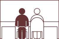

Primary visits take approximately two hours, where as follow-up visits might take around one to one and a half hour. A post operative visit would last for around half an hour.
We accept cash, cheques, net banking, credit card and debit card payments as well as PAYTM.
As a member of Poona Ophthalmological Society we will guide you with the detail information and process at our clinic.
You can find the ‘Eye Donation Pledge Form’ at the following link: ![]() EyeDonationFormPOS.pdf
EyeDonationFormPOS.pdf
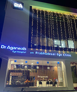
Dr Agarwals Eye Hospital with Prabhudesai Eye Clinic,
Kothrud Branch,
Niksia House Sr no 32/1/1, CTS no 131,
Near Mehendale Garage Chowk, next to Hotel Sweekar,
Erandwane, Kothrud, Pune - 411004, Maharashtra, India.
Landline: +91-9130025349, +91-9130025350
Mobile: +91-9822052063, also for WhatsApp ![]()
For appointments please call between 9:00 am to 8:00 pm
Dr Nitin Prabhudesai is available on
Monday and Tuesday form 9am to 2pm, Wednesday form 2pm to 6pm, Thursday and Friday from 9am to 2pm, and on
Saturday (when available) from 11am to 2pm
Dr Medha Prabhudesai is available from Monday to Saturday from 2pm to 4pm.
Email: contact@prabhudesaieyeclinic.com